Neural Control And Coordination - Class 11 Medical Biology - Extra Questions
Write the function of retina in human eye.
Do you know that the corneal-impairment can be cured by replacing the defective cornea with the cornea of the donated eye? How and why should we organize groups to motivate the community members to donate their eyes after death?
List the parts of eye that controls the amount of light entering in it. Explain how they perform this function.
How does the eye regulate the amount of light that falls on the retina?
Draw labelled diagrams of the following:
(a) Neuron(b) Brain(c) Eye(d) Ear


The names of ten animals whose ears can be seen.
Imagine how it would be to have your eyes In place of your ears? What would you be able to do then, which you cannot do now?
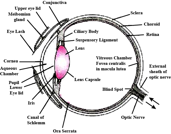
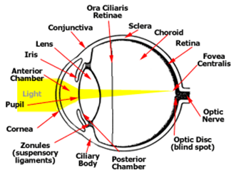
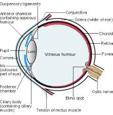
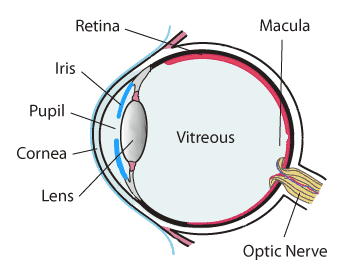
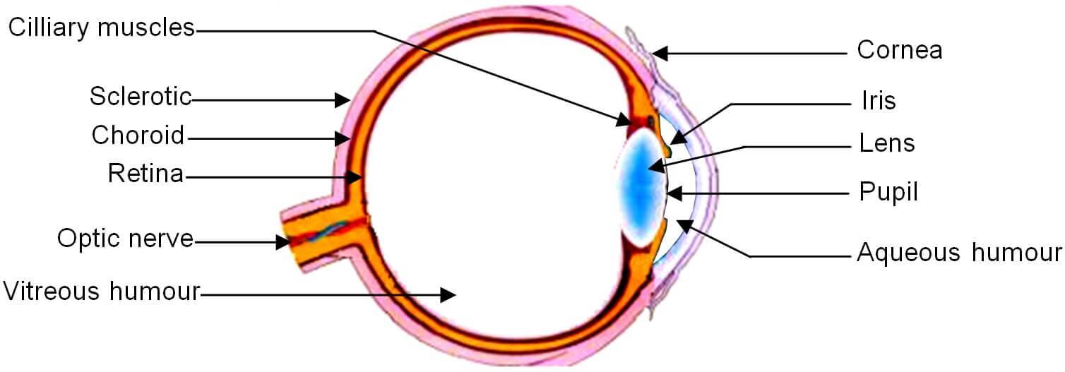
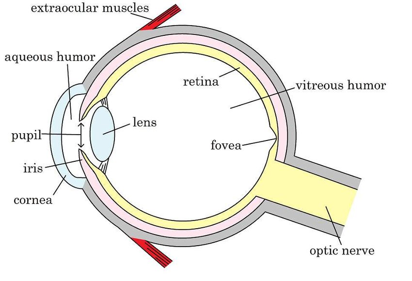
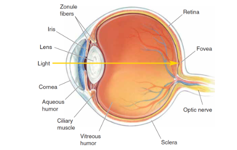
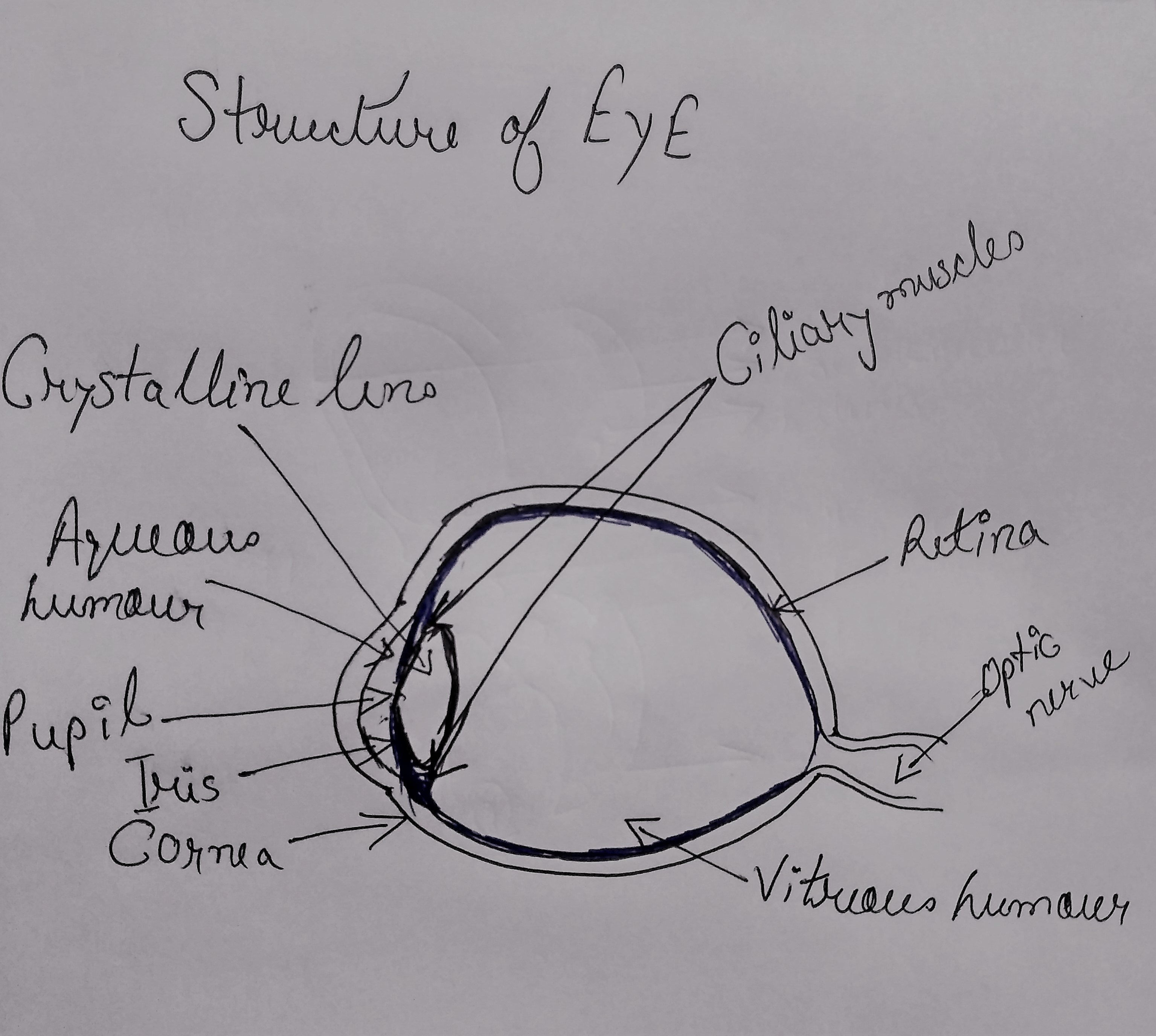
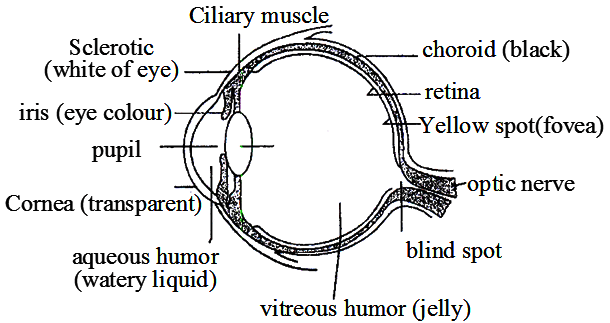
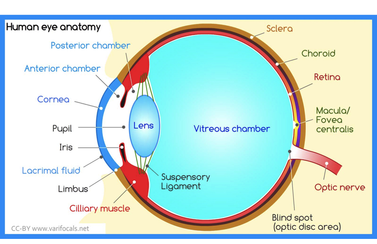
Draw the well labelled diagram of V.S. human eye.
The circular opening enclosed by iris ________.
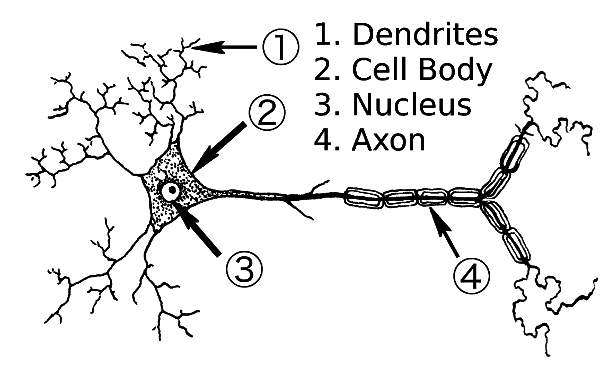
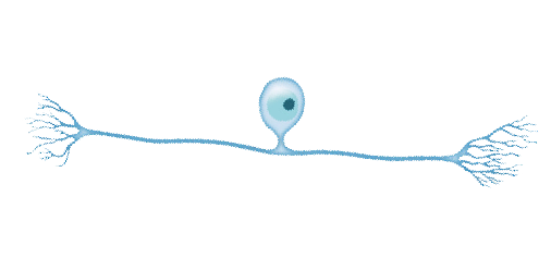
Draw a neat labelled diagram of multipolar neuron.
For this activity find a quiet place in your school. Tell one of your friends to stand at a small distance and ask him to say something softly. The rest of you should listen carefully. Then all of you put your hands behind your ears, as shown in the picture. Let the same child say something again as softly as before. In which case was the sound sharper? Ask our friends too.
Where the tectorial membrane is found?
Write the main parts of the brain.
Sketch and label V. S. of human eye.

Enlist the number and names of motor cranial nerves in a human being ?
Which structures in the eye do not have their own blood supply and therefore rely on diffusion of oxygen and nutrients from adjacent tissue or fluid? What advantage does this avascularity confer?
Spinal nerves are part of which division of the nervous system?
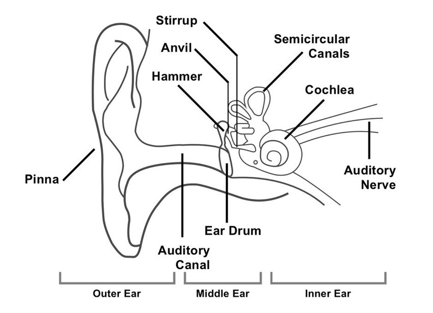
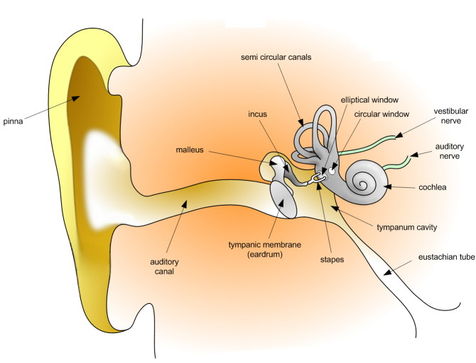
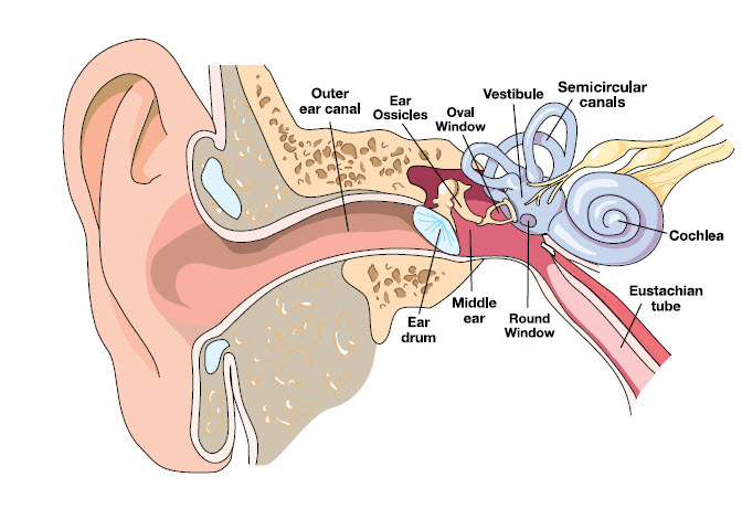
Draw the structure of the human ear and label the following parts :
(i) tympanum (ii) cochlea (iii) pinna (iv) malleus (v) semi-circular canals (vi) eustachian tube
Now can you guess from what distance can an eagle In the sky see a roti on the ground?
State one function of iris in human eye.
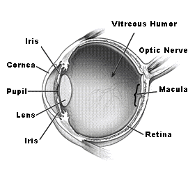
Draw a labelled diagram of V.S. of the human eye and write one function of rod and cone.
(i) Rod : They are responsible for photopic vision and function best at night and dim light.
(ii) Cone : They are responsible for scotopic vision and function in strong light.
Dim light : Rod cells
Bright light:
What are Nissel granules? Where do you find them?
The sympathetic and parasympathetic nervous systems are antagonistic in their actions. Give any two examples in support of this statement.
Write the function of the following part of human eye.
Iris.
Name the enzyme present in tears which prevents eye infections.
Given appropriate biological/ technical terms for the following:
The antiseptic substance in tears.
Draw a labelled diagram of a portion of the sectional view of retina showing its basic structure (no description is required).
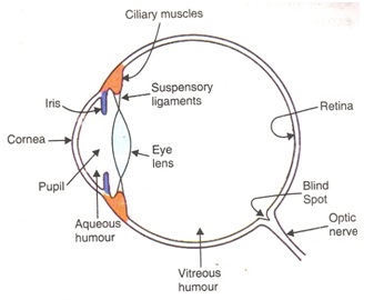
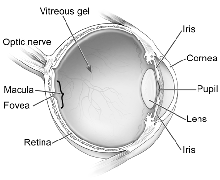
![Illustration of eye anatomy and retinal layers [2], [3]. (a) Cross-sectional view of eye and its major structures. Retina is a thin transparent tissue that lines the back of the eye and is comprised of a number of layers, as illustrated in enlarged portion. (b) Schematic drawing of cellular layers of retina. (a) Two-dimensional illustration of eye anatomy. (b) Schematic of retinal layers. Illustrations from Kolb [3] used with kind permission of Sigma Xi, The Scientific Research Society, Research Triangle Park, NC.](https://www.researchgate.net/profile/Michael_Abramoff/publication/224203356/figure/fig2/AS:670712461275144@1536921720664/Illustration-of-eye-anatomy-and-retinal-layers-2-3-a-Cross-sectional-view-of-eye.jpg)
Draw a neat and labelled diagram of the vertical section of the eye of man and explain its working.

Write short note on retina.
Distinguish between choroid and retina.
Write short note on synapse.
The region of the vertebrate eye, where the optic nerve passes out of the retina, is called as
(a) Fovea(b) Iris(c) Blind spot(d) Optic chaisma
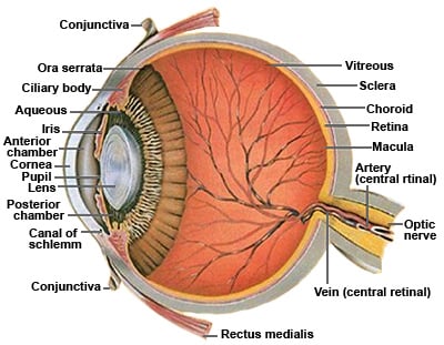
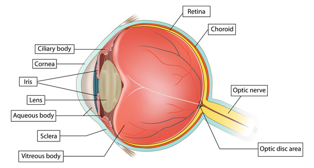
Briefly describe the structure of eye.
Draw a diagram of the human eye as seen in a vertical section and label the parts which suits the following descriptions relating to the :
(i) photosensitive layer of the eye.
(ii) structure which is responsible for holding the eye lens in its position.
(iii) structure which maintains the shape of the eye ball and the area of no vision.
(iv) anterior chamber seen in front of the eye lens.
(v) outer most transparent layer seen in front of the eye ball.
Briefly describe the structure of brain.
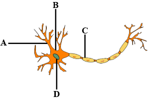
The diagram shows a motor (effector) neurone.
Which structure is also found in white blood cells, but not in red blood cells?
Draw the structure of a neuron and explain its function.
Give reason for tears start flowing while cutting onions.
Find out the false statement and rewrite them as correct one.
Iris patterns are like finger prints used in identifying individuals.
State the difference between pinna and tympanum.
Malleus, Iris, Stapes, Incus.
Fill in the blanks with suitable words. Then give reason why the word is suitable.
Area where optic nerve leaves the eye is called the _________.
Fill in the blanks with suitable words. Then give reason why the word is suitable.
Iris pattern is used for individual ___________.
How would you pay concern towards disabled people who is lacking sensory organs?
State the difference between the two iris and pupil.
Trace the route of sound waves from outer ear to inner ear.
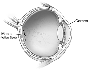
Draw a diagram showing the vertical section of human eyeball and label the following parts.
(a) Cornea
(b) Yellow spot
What is the difference between efferent nerves and afferent nerves?
Write the name of the nerve given in the following diagram and write its function.
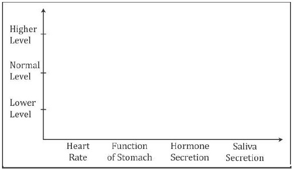
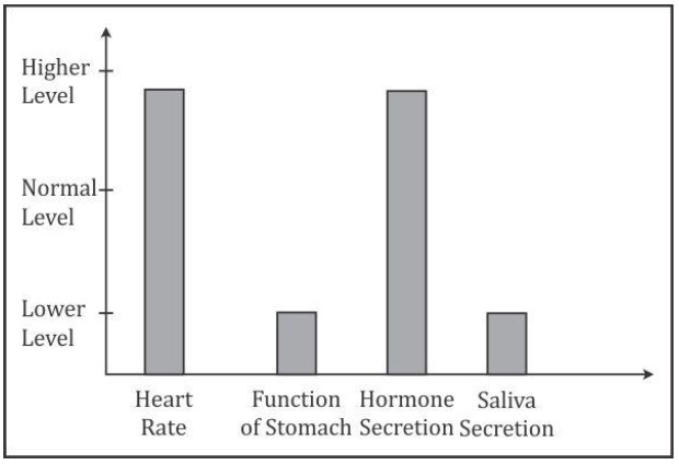
With the help of bar graph illustrated by drawing the functioning of sympathetic system.
Label the following parts.
A. The transparent part of the sclera.
B. The spot on the retina where cones are most abundant.
A. The transparent part of the sclera.
B. The spot on the retina where cones are most abundant.
Redraw the picture.
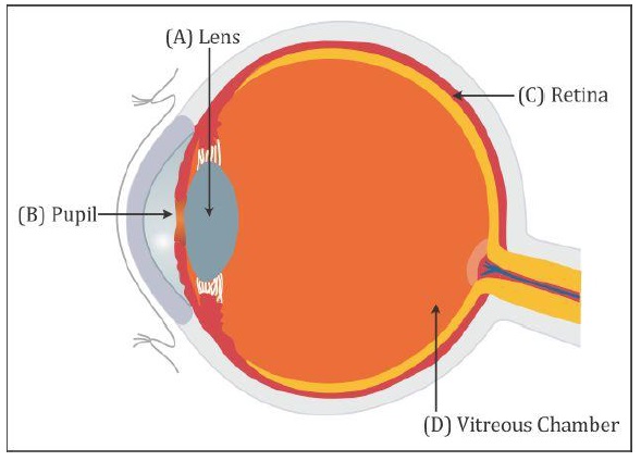
(a) Label the part A, B & C.
(b) Write down the functions of B, C & D.
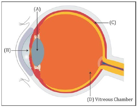
- -Function of B (Pupil)- It regulates the amount of light entering the eye.
- -Function of C (Retina)- It is the site for the formation of image of the objects leading to vision.
- -Function of D (Vitreous Chamber)-The vitreous chamber is the largest cavity in the eye. It helps to maintain the shape of the eyeball.
Name the part of the brain which regulates heart beat and respiration. Where is it located in the brain?
Euglena: .......... :: Insects : Ommatidia
Match the column :
Segregate the appropriate statements from the following as sympathetic and parasympathetic.
- Pupil dilates
- Rate of heartbeat increases
- Secretion of saliva decreases
- Hormone secretion decreases
- Trachea constricts
- Urinary bladder contracts
- Pupil dilates
- Rate of heartbeat increases
- Secretion of saliva decreases
- Hormone secretion decreases
- Trachea constricts
- Urinary bladder contracts
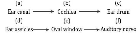
Correct the mistakes and rearrange the given flow chart of hearing process:
Why cornea can be easily transplanted?
Compare the following :
Choroid and Retina.
Answer briefly :
How do you perceive the colour of an object?
What type of striated muscles constitute the muscles of the eyeball? How is this specific type of muscles advantageous?
Which part of the eye has the same name as the Greek goddess of the rainbow.
Mention the function of sympathetic and parasympathetic nervous system with respect to the pupil of eye.
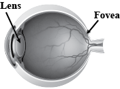
Draw the vertical section of the human eye and label the following parts.
(a) Lens (b) Fovea
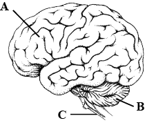
The diagram given below is an external view of the human brain. Study the same and answer the questions that follows:
What are the structural and functional units of the brain? How are the parts of these units arranged in $$A$$ and $$C$$?
What will happen if we have no eyelashes?
Choose the ODD one out of the following terms given and name the CATEGORY to which the others belong:
Aqueous humour, Vitreous humour, Iris, Central canal.
Given below are groups of terms. In each group the first pair indicates the relationship between the two terms. Rewrite and complete the second pair on a similar basis.
Example : Oxygen : Inspiration : : Carbon dioxide : Expiration
Eye : Optic nerve : : Ear : ___________.
Cochlea, Malleus, Pinna, Stapes, Incus.
Choose between the two options to answer the question specified in the brackets for the following.
An example is illustrated.
Example: Corolla or Calyx (Which is the outer whorl?) Answer: Calyx.
Sclerotic layer or choroid layer (Which of the two forms the Iris?)
Write the function of the following part of human eye.
Crystalline lens.
Long Answer Questions
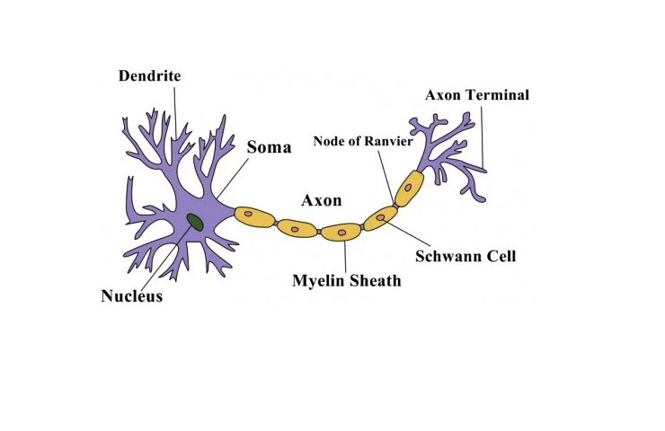
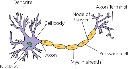
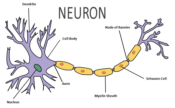
With the help of a suitable diagram describe the structure of a neuron.
The tissue of the nervous system consists of two types of cells, those are the Nerve cells and Glial cell. The nerve cells are called Neuron and the glial cells are called neuralgia. Among these, the neurons performed the function of the nervous system by transmitting information from one part of the body to another and the glial cells are supporting elements.
The Neurons are the structural and functional units of the nervous system.
A typical structure of neuron consists of a cell body and two processes, those are Dendron and Axon. The cell body of a neuron is like other cells of the body, consisting of a nucleus and cytoplasm covered by a plasma membrane. The cytoplasm contains various organelles and inclusions. In adult nerve cells, the centrosome is absent or inactive so their division does not occur. Specialized cytoplasmic organelles called Nissl granules or Nissl body made up of ribonucleoprotein are found in the cell body except in the axon hillock region from where the axon arises. These granules are also present in dendrons but absent in axon.
In general, a neuron has many dendrons. However, in some neurons, the Dendron may be only single or may be absent in case. The single long efferent process of a neuron is called axon which carries impulses away from the cell body to the next neuron or the target organ, such as muscle or gland.
Axons are usually unbranched. However, they give a few small terminal branches called telodendria having swollen ends known as terminal buttons. An axon is also called nerve fiber. A neuron possesses only one axon and the variable number of dendrons. If there is only one process, it is usually the axon. In such type of neurons, the cell body receives stimulation. In some specialized neurons, the axon may be absent, such as adrenal medullary cells and amacrine cells of a retina of an eye.
Describe three kinds of nerves, giving example of each.
Name the layer of the eyeball that forms the transparent cornea.
Mention the location and function of given parts:
(a) Fovea centralis
(b) Choroid
Complete the following statements by choosing the correct alternative from the choices given in brackets
(a) The dorsal root ganglion of the spinal cord contains cell bodies of (motor/sensory/Intermediate) neurons.
(b) Cerebellum is the part of the brain which is responsible for
(i) conducting reflexes in the body
(ii) maintaining posture and equilibrium
(iii) controlling thinking, memory & reasoning.
(c) Reflex action is controlled by
(i) brain
(ii) spinal cord
(iii) autonomic
(iv) peripheral nervous system.
Classify the following as voluntary actions and involuntary actions.
Sneezing, throwing ball, heart beats, writing.
Rearrange the following in the correct sequence pertaining to what is given within brackets at the end.
(a) Effector Sensory neuron Receptor Motor neuron Stimulus Central nervous system Response (Reflex arc)
(b) Repolarisation Depolarisation Resting (polarised) (during conduction of nerve impulse through a nerve fibre)
(c) Axon endings Nucleus Dendrites Axon Perikaryon Dendron (Neuron structure)
(d) Diencephalon Cerebellum Medulla oblongata Pons Cerebrum mid brain (sequence of parts of human brain).
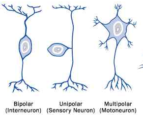
Name different types of neurons and give one function of each.
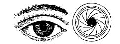
The figure below compares a part of our eye with a part of a photographic camera.
(a) Name the corresponding parts of the eye and the camera shown here that are comparable in function.
(b) Explain the mode of working and the functions of the parts of the eye mentioned above.
Very short answer type.
Which are the neurons found mostly in the central nervous system?
With reference to the human ear, answer the questions.
(a) Give the technical term for the structures found in the inner ear.
(b) Name the three small bones present in the middle ear. What is the biological tam for them collectively?
(c) Name the part of the ear associated with (1) static balance (2) heating (3) dynamic balance.
(d) Name the nerve, which transmits messages from the ear to the brain.
Fill in the blanks with appropriate words.
A neuron receives a nerve impulse through _______and transmits it through ________.
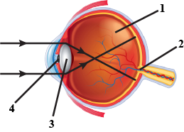
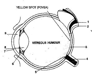
Given is a diagram depicting a defect of the human eye. Study the same and answer the questions that follow:
(a) Name the defect shown in the diagram.
(b) Give two possible reasons for this defect.
(c) Name the paws labeled 1 to 4.
(d) Name the type of lens used to correct this eye defect.
(e) Draw a labelled diagram to show how the above mentioned defect is rectified using the lens named above.

.......... receive information from sensory receptors and transmit it to the central nervous system.
Fill in the blanks with appropriate words.
Vertebrate nervous systems are divided into two major divisions the central nervous system, which includes the .......... , and .......... and the .......... nervous system, which includes all neurons other than those in the brain and spinal cord
Which muscles rolls the eyeballs laterally?
Name a cranial nerve that controls eye ball movements. Is this nerve a sensory, a motor or a mixed nerve?
Very short answer type.
Name the area of the retina which has the brightest vision.
Name the neuron which is responsible for one-way impulse conduction.
Name the ear ossicles in the order of arrangement in humans.
Name the area of the retina which contains only cones and no rods.
Sound moves through an ear when pressure waves in the .......... cause vibrations of the .......... membrane.
Make a labelled diagram of a vertical section of a mammalian eye. Describe what happens when one looks from a distant object to a near one. Explain the functions of rods and cones
Very short answer type.
What are tears, and what are their functions?
Draw a vertical section of the human eye and label any six of those parts only through which the light rays pass and fall on the retina.
Draw a diagram of the vertical section of the human eye and label the following parts: (i) cornea (ii) iris (iii) aqueous humour (iv)retina (v) ciliary body (vi) fovea centralis (vii) choroid (viii) pupil (ix) lens

What are the two divisions of the autonomic nervous system? Show by taking an example, how these divisions are antagonistic in their actions. Explain with reference to any three organs in the human body.
What are the functional similarities between rods and cones?
Draw a diagram of the vertical section of human eye and label any six parts surrounding the vitreous chamber.

Differentiate between.
Motor neurons and sensory neurons
Short / Long answer type question:
Which division- sympathetic or parasympathetic of the autonomic nervous system has longer preganglionic fibres and why?
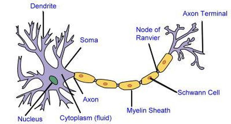
Draw a well labelled diagram of a non-myelinated neuron.
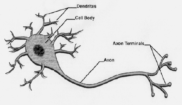
Draw neat labelled diagram of neuron.
What are sensory and motor neurons? Write their functions.
Name the following:
Kind of retinal cells sensitive to dim light
The structure responsible for protecting the eye from sweat is ............
Name the following:
Capacity of the eye to focus at different distance
Place of best vision in the retina of the eye is _________.
Write in proper sequence the name of all the parts of the human eye through which the light rays coming from an object pass before they form an image on the retina.
The internal layer of the eye which prevents reflection of light is known as ............
Name the following:
The fibres which collectively hold the lens in position
Name the following:
The layer of the wall of the eye-ball that corresponds to the black lining of the bix of a camera
Describe, giving diagram, the structure of the eye and explain the process vision.
Define associative neuron.
Some neurons have a __________ around their axons.
Distinguish between afferent and efferent neurons?
The part of the human body which is called as second brain, is _____
We feel blinded for a short while entering a dark room when coming from bright light.
The projections arising from the cell body are?
Sympathetic nervous system is also called?
Long Answer Question:
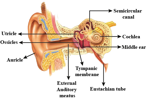
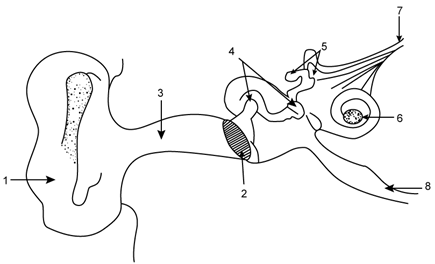
Describe the internal structure of human ear.
- The ear performs two sensory functions, hearing and maintenance of body balance.
- Anatomically, the ear can be divided into three major sections called the outer ear, the middle ear, and the inner ear.
- The vestibular apparatus is composed of three semicircular canals and otolith organ consisting of the saccule and utricle.
Give the role of following : (a) Medulla oblongata (b) Cerebellum.
Neural system and computers share certain common features. Comment in five lines. (Hint: CPU, Input-output devices).
Name and write one point of difference each between the two kinds of Autonomic Nervous system with respect to their effects on
(a) Heart
(b) Urinary Bladder.
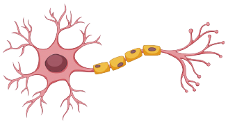
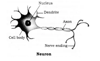
What does a neuron look like?
Long Answer Question:
Describe different part of human eye?
Draw the structure & functions of neurons.

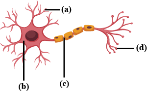
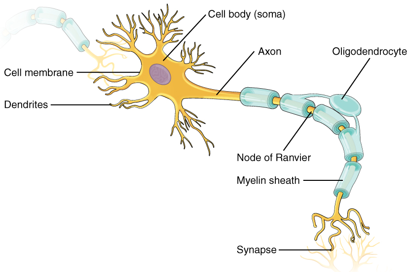
The structure of a neuron: The above image shows the basic structural components of an average neuron, including the dendrite, cell body, nucleus, Node of Ranvier, myelin sheath, Schwann cell, and axon terminal.
Function of a Neuron
The specialized structure and organization of neurons allows them to transmit signals in the form of electric impulses from the brain to the body and back. Individually, neurons can pass a signal all the way from their own dendrites to their own axon terminals; but at a higher level neurons are organized in long chains, allowing them to pass signals very quickly from one to the other. One neuron’s axon will connect chemically to another neuron’s dendrite at the synapse between them. Electrically charged chemicals flow from the first neuron’s axon to the second neuron’s dendrite, and that signal will then flow from the second neuron’s dendrite, down its axon, across a synapse, into a third neuron’s dendrites, and so on.
Draw the structure of neuron & explain its functions.
What is the longest cell in the human body?
What is the name of the part of a human eye where image of an object is formed?
State the function of each of the following parts of the human eye;-
(a) Cornea
(b) Iris
(b) Iris
State the function of each of the following parts of human eye ;-
(a)Cornea
(b)Iris
What do grey and white matter in the brain represent?
Give an account of the retina of the human eye.
Why is the sympathetic division called thoracolumbar division?
Match the following
1) Distilled a) Carries hereditary
2) Budding b) In between 20Hzto to 20000Hz
3) Audible range of human ear c) Poor conductor
4) Chromosome d) Cell
5) Basic living unit e) Yeast
1) Distilled a) Carries hereditary
2) Budding b) In between 20Hzto to 20000Hz
3) Audible range of human ear c) Poor conductor
4) Chromosome d) Cell
5) Basic living unit e) Yeast
Give biological reason for the following:
One feels blinded for a short time while coming out of a dark room.
The gap between dendrites of one neuron and axon endings of other neurons.
Structure which is responsible for holding the eye lens in its position.
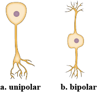
Copy and identify the types of neurons given below:
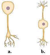
- A unipolar neuron is a type of neuron in which only one protoplasmic process (neurite) extends from the cell body.
- A bipolar neuron or bipolar cell is a type of neuron which has two extensions (one axon and one dendrite).
Structure which maintains the shape of the eye ball and the area of no vision.
What happens at the synapse between two neurons?
Draw a well labelled diagram of human eye and write functions of following parts: retina, pupil, ciliary muscles, and iris.
The ear ossicle in contact with the oval window.
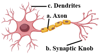
Labels the parts of a neurons in figure.
Analyse the following information and answer the questions.
| SI. No. | Organ | List-1 Effect of Nervous system | List-2 Effect of Nervous system |
| 1 | Eye | Dilates pupil | Constricts pupil |
| 2 | Mouth | Inhibits salivation | Stimulates salivation |
| 3 | Lungs | Relaxes bronchi | Constrict bronchi |
| 4 | Heart | Accelerates heartbeat | heartbeat to normalcy |
| 5 | Blood vessel | Increase blood pressure | Decrease blood pressure |
| 6 | Pancreas | Inhibits pancreas activity | Stimulates pancreas activity |
Name the neurons which carry impulses from receptor to brain.
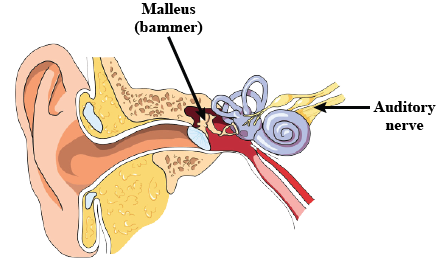
Draw the diagram showing the internal structure of human ear and label the following parts
(i) Malleus (ii) Auditory nerve
Analyse the following information and answer the questions.
| SI. No. | Organ | List-1 Effect of Nervous system | List-2 Effect of Nervous system |
| 1 | Eye | Dilates pupil | Constricts pupil. |
| 2 | Mouth | Inhibits salivation | Stimulates salivation |
| 3 | Lungs | Relaxes bronchi | Constrict bronchi |
| 4 | Heart | Accelerates heartbeat | heartbeat to normalcy |
| 5 | Blood vessel | Increase blood pressure | Decrease blood pressure. |
| 6 | Pancreas | Inhibits Pancreas activity | Stimulates Pancreas activity |
Redraw the diagram. Name and label the parts indicated below
a. Carries impulses away from the cell body
b. Secretes neurotransmitter
c. Branches of dendrons
Describe the structure of medulla oblongata.
What is autonomous nervous system?
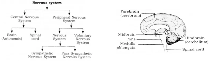
Describe with the help of diagram, the structure of the human nervous system and functions of the brain.
(a) The central nervous system.
(b) The peripheral nervous system.
(a) The Central Nervous System: It consists of the brain and spinal cord. It is responsible for the coordination and control of the activity of the nervous system. CNS enables a person to give a more appropriate and more intelligent response to various situations. The work of the central nervous system is to collect all the information from all the receptors in our body. In complicated responses the brain and spinal cord, both are involved.
1. Brain: Human brain is shaped like a large mushroom. It is located in the bony cavity of the skull, called cranium. The brain is covered by three membranes, called meninges. The fluid (cerebrospinal) is filled between these membranes. This fluid protects the brain from mechanical shocks.
The brain is divided into three regions:
(a) Forebrain,
(b) Midbrain,
(c) Hindbrain.
The forebrain is the largest region, covering 2/3 portion of the brain. It mainly consists of the cerebrum. Midbrain is quite small and inconspicuous. It connects cerebrum to the other parts of the brain and spinal cord.
Hindbrain consists of three main parts
(a) medulla oblongata,
(b) pons and
(c) cerebellum.
2. Spinal cord: It consists of a number of nerve cells, both fibres and cell bodies, grouped into a cylindrical mass that extends from the brain to the bottom edge of the first lumbar vertebra. It is covered by the same meninges as the brain and lies within the neural canal of the vertebral column. A cross section of the spinal cord reveals an outer region of white matter and an inner H-shaped region of grey matter. The grey matter consists of cell bodies of thousands of neurons.
The white matter is composed of myelinated nerve fibres, running up and down the cord or passing out to the spinal nerves. Running through the centre of the spinal cord is the central canal. Dorsal and ventral fissures (grooves), divides spinal cord into right and left halves. The spinal cord has two-fold
functions: It conducts impulses to and from the brain and acts as a reflex centre.
(b) Peripheral nervous system: This consists of three parts: Cranial nerves; Spinal nerves and Autonomic nervous system.
Cranial nerves: The cranial nerves are those that emerge from the brain e.g. in man, there are twelve pairs of cranial nerves, which are mainly supplied by the organs in the head region.
Spinal nerves: These are supplied by the spinal cord. There are 31 pairs of spinal nerves, all of which are mixed nerves. These include 8 cervical 12 thoracic, 5 lumbar, 5 sacral and 1 coccygeal.
Autonomic nervous system: This lies outside a.nd parallel to the central nervous system and is organised into two distinct regions, sympathetic system and parasympathetic system. While the former consists of thoracic and lumbar ganglia and using only spinal nerves, the latter uses three spinal nerves of the lower region of the cord, i.e., sacral part and four cranial nerves. The autonomic nervous system consists of two sets of nerves with opposite actions- If one speeds up activity, the other decreases it. The autonomic nervous system controls the functions of the internal organs of the body automatically and unconsciously.
It sends impulses via the efferent or motor neurons to the effector structures, other than the skeletal muscles. These structures include smooth muscles, lining the wall of digestive, respiratory, urinary and reproductive tracts; the smooth muscles of blood vessels and eye, the cardiac muscles of heart and both, exocrine and endocrine glands. Thus, the autonomic nervous system regulates
1. the contraction of the involuntary muscle,
2. the rate and force of the heartbeat,
3. the secretion of the glands of the alimentary tract, and
4. the size of the pupil of the eye.
Functions of Brain:
1. The brain receives impulses from all sensory organs, such as eyes, ear, etc.
2. After receiving signals or impulses from sensory organs, the brain sends off instructions to muscles and glands, to respond through motor neurons.
3. Brain correlates various stimuli from different sense organs.
4. Brain efficiently coordinates the activities of our body.
5. Brain stores information, in the form of our knowledge.
6. The brain is responsible for thinking and intelligence.
What is somatic nervous system?
Complete the paragraph.
The milk was on the stove. Rasika was engrossed watching television. She smelled something burning. She ran towards the kitchen. The milk was boiling over. She held the vessel with her bare hands but, screaming, she let it go at once. This activity was controlled by _____ cells. Special ends of _______ in these cells collected the information, from where it was transferred to the ______ and then towards the terminal end of the ______ The chemicals produced at the terminal end passed through the minute space i.e. ______. In this way, ________ were conducted in the body and the process of ________ was completed by conducting the impulses from _______ to _______
(Nerve, Muscle cell, Impulse, Dendrite, Synapse, Axon, Reflex action, Cell body)
Which cells of the retina enable us to see coloured objects around us?
Our reaction like aggressive behavior. use of abusive words, restlessness etc. are regulated by brain, name the parts involved.
Write a short note on:
Nervous tissue
1. Cyton: It is the cell body of a nerve cell, that has a large central nucleus and cytoplasm, from which long thin hair-like extensions arise.
2. Dendrite: It is the short branched fibre of neuron, which receives nerve impulses.
3. Axon: It is the single long conducting fibre extending from a neuron, that transmits impulses away from the cell body.
Name the principal body heat regulating centre in our brain.
Why the name peripheral nervous system is given?
Which are two parts of the autonomous nervous system?
What is the function of the sensory division of the peripheral nervous system?
Draw a well labelled diagram of eye.
State the exact location of the following structures.
Ciliary body.
Neurons get nutrients from supportive cells called as
Match these parts with their functions:(Medulla oblongata, cerebellum, forebrain, thalamus, cerebral cortex, hindbrain, pons, hypothalamus)
a) Sleep centre and respiratory centre.
b) Several reflexes involved in the regulation of heartbeat, blood vessel contraction, breathing, etc.
c) Consists of cerebrum, thalamus, and hypothalamus.
d) Motor and sensory areas.
e) A major conducting centre for sensory and motor signaling.
f) Regulation of sexual behavior.
g) Consists of the pons, cerebellum, and medulla oblongata.
h) Coordinates the group movements of voluntary muscles, as in walking or running.
Draw a diagram of vertical section of human eye and label the following parts.
a) Fovea
b) Lens
Describe the structure of neuron with the help of a neat labelled diagram.
Name any 5 types of nerve cells.
Describe the structure of human eye.
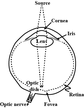
Draw a line sketch to show the image formation on the retina of human eye.
With the help of a neat and labelled diagram describe the anatomy of human eye. Explain the mechanism of vision.
Name six parts or regions of the eye through which light must pass before reaching the retina.
Draw a neat and labelled diagram of structure of the human eye.
The diagram given above represents a vertical section through the human eye. Name the parts numbered 1 to 9
Fill in the blanks with appropriate words.
A neuron receives a nerve impulse through .......... and transmits it through ..........
Differentiate between.
Neuron and nephron.
Difference between the dendrites and axons.
Among thalamus, hypothalamus, cerebral cortex, cerebral medulla, cerebellar cortex, cerebellar medulla, pons and medulla, which are composed of grey matter and which are with white Matter? Support your answer with the reason
Where are neurons found in the body and what is their function?
(a) The figure shows the principal parts of the human ear. Study the diagram and answer the following questions:
(i) Label the parts 1-8.
(ii) State the role of parts 6 and 7.
(iii) Why is it harmful to use a sharp object to remove wax from our ears?
Class 11 Medical Biology Extra Questions
- Anatomy Of Flowering Plants Extra Questions
- Animal Kingdom Extra Questions
- Biological Classification Extra Questions
- Biomolecules Extra Questions
- Body Fluids And Circulation Extra Questions
- Breathing And Exchange Of Gases Extra Questions
- Cell Cycle And Cell Division Extra Questions
- Cell The Unit Of Life Extra Questions
- Chemical Coordination And Integration Extra Questions
- Digestion And Absorption Extra Questions
- Excretory Products And Their Elimination Extra Questions
- Locomotion And Movement Extra Questions
- Mineral Nutrition Extra Questions
- Morphology Of Flowering Plants Extra Questions
- Neural Control And Coordination Extra Questions
- Photosynthesis In Higher Plants Extra Questions
- Plant Growth And Development Extra Questions
- Plant Kingdom Extra Questions
- Respiration In Plants Extra Questions
- Structural Organisation In Animals Extra Questions
- The Living World Extra Questions
- Transport In Plants Extra Questions